Life Sciences Imaging Facility Provides access, support and training in advanced light and fluorescence microscopy techniques. Contact Us
Start main page content

Life Sciences Imaging Facility

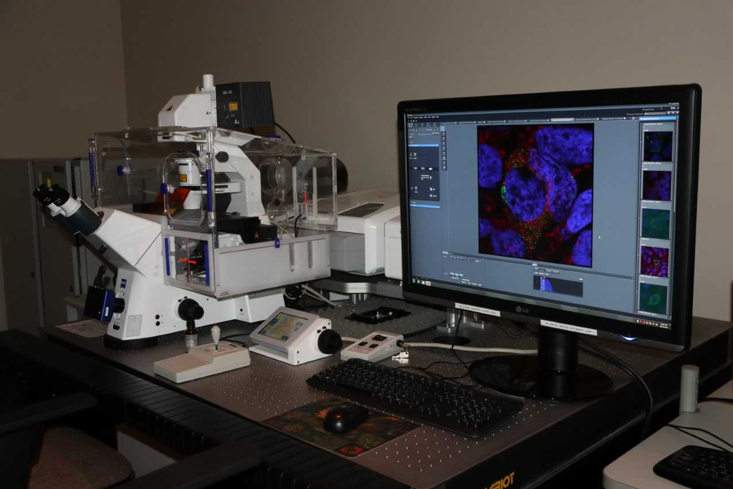
The cross Faculty (Science and Health Sciences) Life Sciences Imaging Centre is an open central facility located at the Medical School of the University of the Witwatersrand.

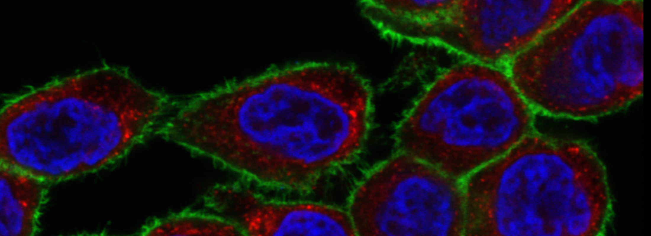
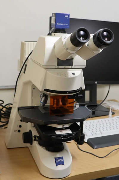
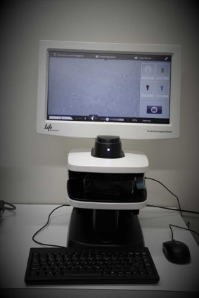
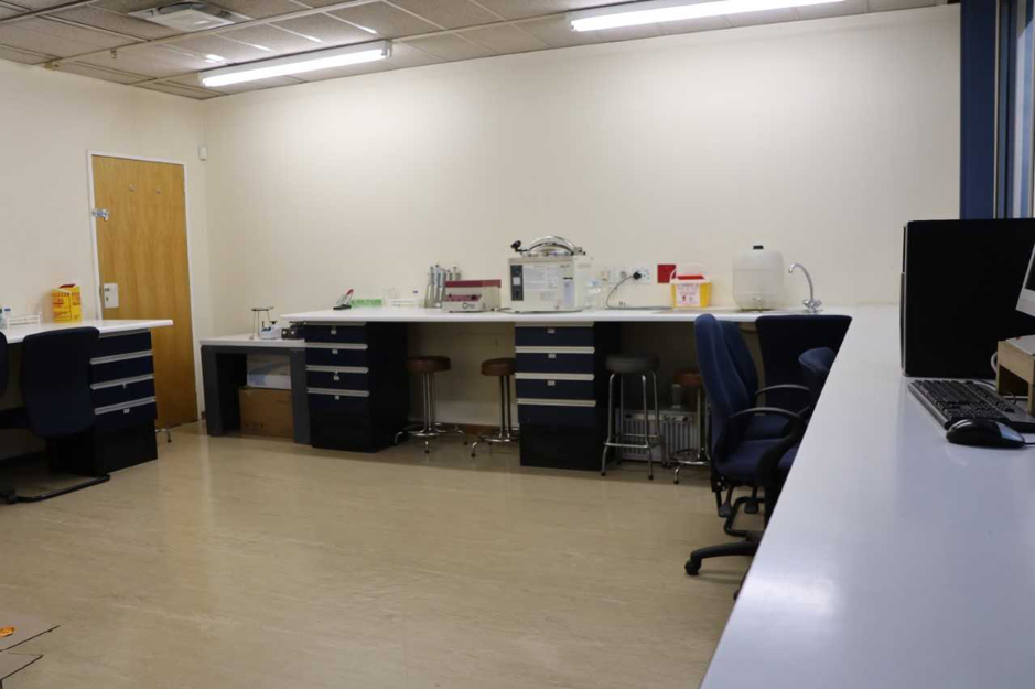
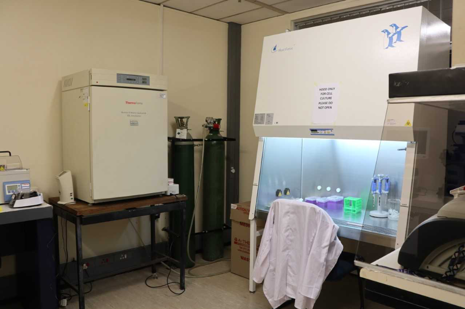
Our mission is to develop a teaching and learning centre, to assist Honours, Masters and Doctoral students, post-doctoral fellows and all staff members with microscopy related research. We provide services to develop molecular and cell biology studies at Wits with a cell culture room, a preparation room for biological samples and our confocal and bright-field/fluorescence microscopes. The objective of the Centre is to provide current and emerging technologies involving microscopy and imaging in Life Sciences. We are promoting cutting edge research in basic and applied sciences, which include a wide range of studies: viruses, plants, bacteria, fungi, pharmacology, genetics and neurosciences are some examples. We promote quality training and education through individual training sessions, lectures for students participating in different Life Sciences programs and formal courses organized with our different partners, such as Carl Zeiss.

Services offered:

  • Individual instruction in the use of the confocal microscope for students and all staff members
  • Academic assistance with project design, cell culture, sample preparation, image capturing and image analysis
  • Workshops organized in the microscopy unit during the year.
  • Co-supervision of students
Zeiss Laser Scanning Confocal Microscope (LSM) 780 and Airyscan

Confocal microscope scans samples sequentially point by point, or multiple points at once. The pixel information is assembled into an image. As a result, you acquire optical sections with high contrast and high resolution in x, y and z.

  • Lasers:
  • Uni‐ and bidirectonal scan, simultaneous and/or sequential image acquisition
  • Live cell imaging
  • Differential Interference Contrast (DIC)
  • 3D imaging with z-stack acquisition
  • Multiple time series
  • FRET, FRAP
  • Linear unmixing

The Airyscan is a ground-breaking new add-on unit that transforms a standard confocal microscope into a super-resolution microscope with 1.7x higher resolution in all three dimensions.  Unlike other super-resolution techniques, the Airyscan is not restricted by complicated biochemical labeling procedures or dye selections. With Airyscan, you collect all precious emission light without compromise and get a unique combination of gentle supperesolution imaging and high speed. You can get resolved structures of 120nm (in x and y) and 350nm (in z). It gives you an instant 4-8X improvement of signal-to-noise ratio.

Zeiss Fluorescent Microscope- Axioskop 2

Upright microscope with all the standard microscopy technique- from transmitted light brightfield to epi-fluorescence. You can take pictures with its new Axiocam Icm 1 camera.

Floid Cell Imaging Station (ThermoFisher)

For a quick detection and verification of fluorescently-labeled sample, the Floid captures transmitted light and three-color fluorescent images of your cells and samples.

Staining Laboratory

Designed for students to proceed with immunofluorescence and for our microscopy workshops, the lab can host up to 16 students.

Cell Culture Room

This room is very useful for students coming from outside Medical School who have to do live cell imaging. Leave your sample in the incubator and carry on with confocal microscope.

Thermal Cycler (G-Storm, Vacutec)

Thermal Block Characteristics

  • Temperature Range- 4°C - 99°C
  • Volume Range - 2-150μl
  • Ramp Rate - (standard block) Up to 3.8°CS-1 / (fast block) up to 6ºCS-1
  • Block Accuracy - +/- 0.2°C (20-99°C)
  • Gradient Temperature Range - 30ºC - 80ºC

Share